
Vascular and Biliary Variants in the Liver Implications for Liver Surgery RadioGraphics
The level set segmentation uses an initial user-defined liver segment in one slice, and then segments the liver through all other slices, using a Gaussian fit to define the speed image where the level sets propagates.. Kuni O. Liver CT image processing: A short introduction of the technical elements. European Journal of Radiology. 2006; 58:.
The Radiology Assistant Liver Segmental anatomy
We aim to develop and validate a three-dimensional convolutional neural network (3D-CNN) model for automatic liver segment segmentation on MRI images. Methods This retrospective study evaluated an automated method using a deep neural network that was trained, validated, and tested with 367, 157, and 158 portal venous phase MR images, respectively.

Couinaud classification of hepatic segments Radiology Reference Article
This article segments liver and liver tumor CT scans using ResU-Net. The localization results demonstrate that the proposed method more accurately localized the very minute liver tumor. The Liver CT scan segmentation is quantitatively evaluated in terms of DSC, accuracy, precision, specificity, VOE, and RVD values averaged over all test images.
The Radiology Assistant Liver Segmental anatomy
CT CHEST by Raeesa Kabir; Ct lung segments annotated by Karl Fan; Annotated Anatomy by Neagu Andrei; UQ BIOM3002 - Chest by Craig Hacking Mohit Ct by Mohit Seth; ct by keerthana s; UQ Radiologic Anatomy 4. Chest 4.2 Tracheobronchial Tree by Craig Hacking UQ Radiologic Anatomy 4. Chest 4.3 Pleura by Craig Hacking مهم by majid alipoor
The Radiology Assistant Liver Segmental anatomy
Dataset. The 3D-IRCADb-01 database is composed of three-dimensional (3D) CT-scans of 20 different patients (10 females and 10 males), with hepatic tumors in 15 of those cases. Each image has a resolution of 512 × 512 width and height. The depth or the number of slices per patient ranges between 74 and 260.
Radiopaedia CTscan Hepatic segments coronal section labels AnatomyTOOL
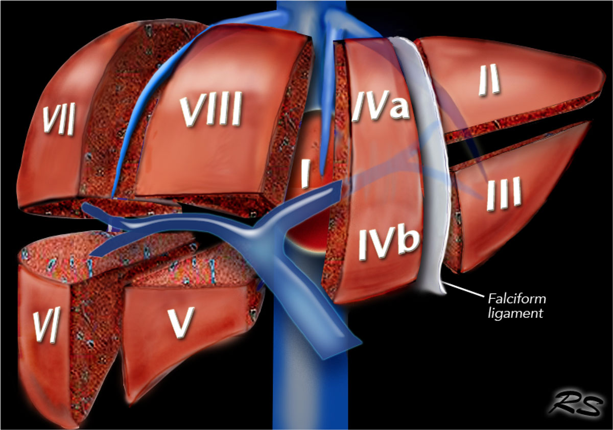
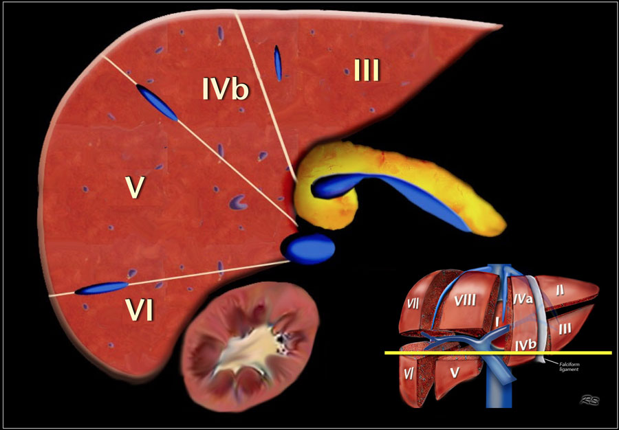
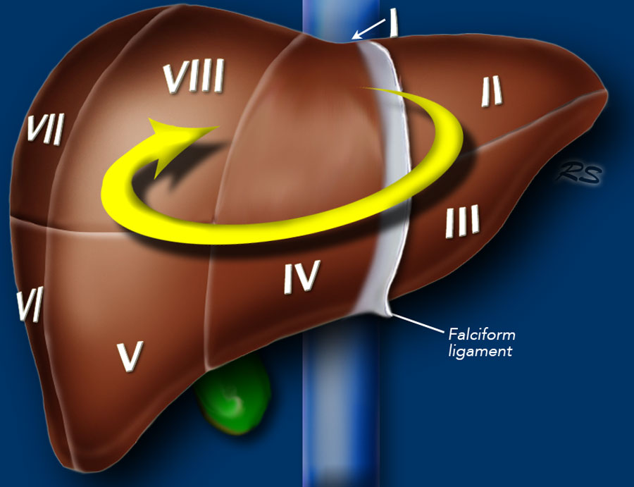
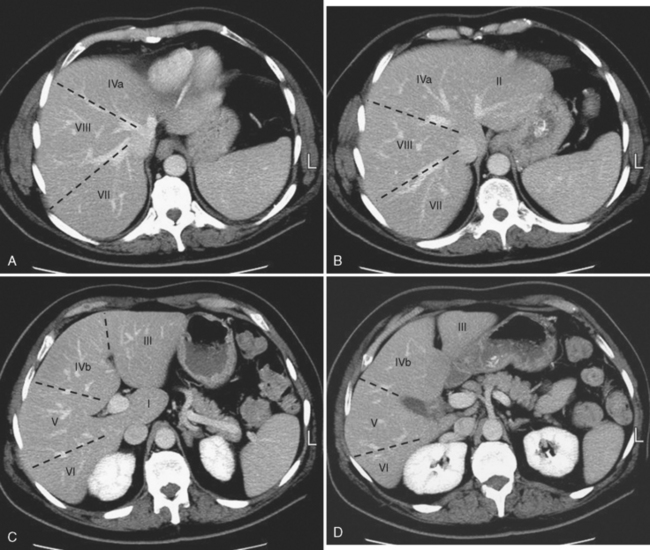
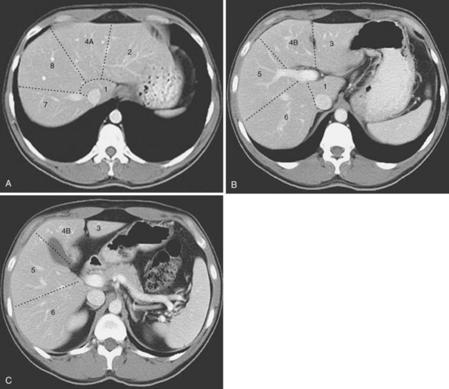
Hepatic segments. Each sector is subsequently divided into two, producing eight hepatic segments. If the patient is supine, and the liver is reflected along its inferior border towards the diaphragm, the segments would be numbered in an anti-clockwise manner around the porta hepatis.. Segment I - the caudate lobe - is the posterosuperior part of the left medial sector.
Liver Anatomy Segments Anatomical Charts & Posters
This work presented the development of an automatic method for liver and tumor segmentation from CT scans. The proposed method was based on fully convolutional neural (FCN) network with region-based level set function. The framework starts to segment the liver organ from CT scan, which is followed by a step to segment tumors inside the liver.
The Radiology Assistant Liver Segmental anatomy
Segmenting a liver and its peripherals from abdominal computed tomography is a crucial step toward computer aided diagnosis and therapeutic intervention. Despite the recent advances in computing.
Liver Anatomy Segments Anatomical Charts & Posters
Definition The hepatic segmentation (lobes, parts, divisions and segments) is the oganization of the liver into parts, divisions and segments.
Liver Radiology Key
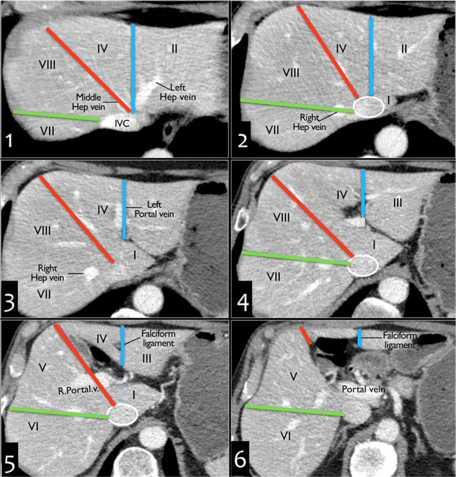
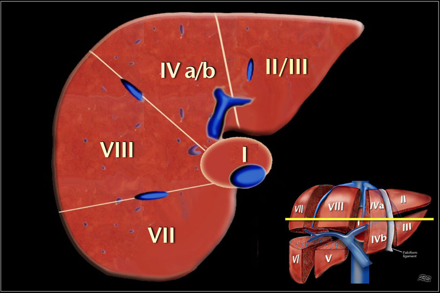
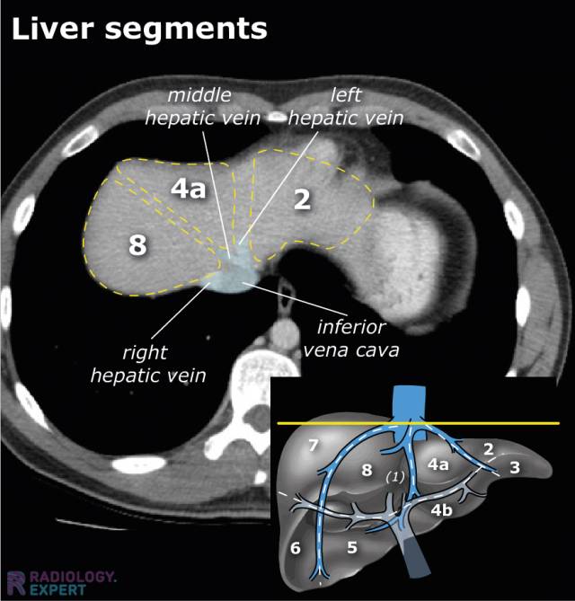
Segmental anatomy according to Couinaud. Click to enlarge. Couinaud classification The Couinaud classification of liver anatomy divides the liver into eight functionally indepedent segments. Each segment has its own vascular inflow, outflow and biliary drainage.

Anatomy of the liver segments Liver anatomy, Medical radiography, Medical ultrasound
Finally, the Couinaud anatomical segments are identified according to the anatomical liver model proposed by Couinaud. Results: Experiments were conducted using data and metrics brought from the liver segmentation competition held in the Sliver07 conference. A subset of five exams was used for estimation of segmentation parameter values, while.

Segmental anatomy of liver segments 18 of liver as depicted on... Download Scientific Diagram
segment 1 (I) is the caudate lobe bounded posterolaterally by the fossa for the inferior vena cava, anteriorly by the ligamentum venosum, and inferiorly by the porta hepatis its inferior portion is subdivided into a lateral caudate process and a medial papillary process 6 may receive its supply from both the right and the left portal vein
Liver Radiology Key
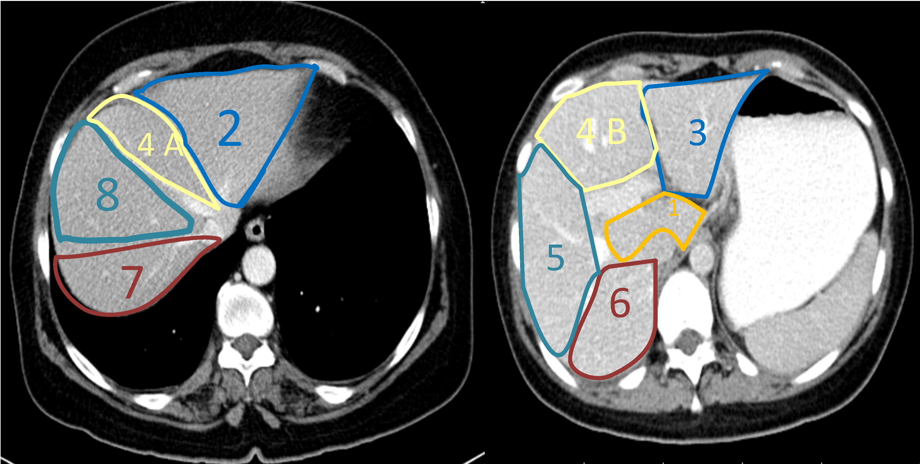
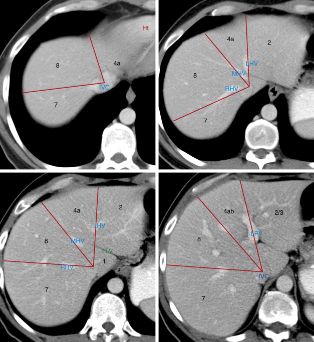
Gender: Female. Annotated image. Annotated Axial CT with contrast of hepatic segmentation and veins.

Liver segments annotated CT Radiology Case Liver anatomy, Radiology
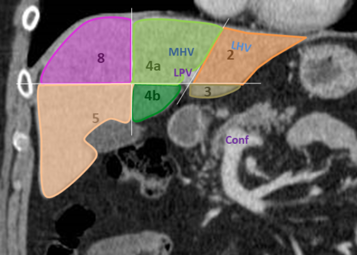
Annotated image. Annotated Coronal CT showing hepatic segmentation and veins. IVC = Inferior Vena Cava. RHV = Right Hepatic Vein. MHV = Middle Hepatic Vein. LHV = Left Hepatic Vein. PV = (Main) Portal Vein. RPV = Right Portal Vein. LPV = Left Portal Vein.
CT abdomen general
Fully Automated Measurements of Liver Segments and Spleen Volume. Two DL models developed in-house were used to automatically segment the eight liver Couinaud segments and spleen from a CT volume. Details of the training data and model development are provided in Appendix E1 (supplement). The outputs of the models include the segmentation.
The Liver Thoracic Key
Each segment has its own pulmonary arterial branch and thus, the bronchopulmonary segment is a portion of lung supplied by its own bronchus and artery. Each segment is functionally and anatomically discrete allowing a single segment to be surgically resected without affecting its neighboring segments. There is some form of segmental symmetry.